Ever thought about how a body scan could turn a fuzzy image into one that's crystal clear? Modern medical imaging goes way past old-school X-rays by giving doctors a peek at even the tiniest details inside our tissues. It's like swapping out a blurry photo for a sharp, vivid picture, which helps doctors decide faster and care better. With safer, quicker scans, patients really benefit. Let's take a closer look at how these new techniques are making a big difference in patient care.
Medical Imaging Advances: Modern Breakthroughs and Clinical Significance
Medical imaging has come a long way. We once relied on simple X-rays and ultrasounds, but now we have advanced optical systems that let us see tiny details in our tissues and organs. It’s like switching from a fuzzy snapshot to a high-definition movie, turning ordinary scans into powerful tools for diagnosis.
Digital technology has completely changed how we capture and share images. Digital mammography, for example, gives doctors quick, sharp pictures that help catch problems early. And with digital radiography, medical teams can share images almost instantly, speeding up decisions and improving patient care. These changes are part of a bigger flood of medical breakthroughs that deliver accurate, timely information straight to the clinicians.
- Digital mammography improvements – Fast, clear pictures help catch issues sooner.
- Minimally invasive CT protocols – New CT methods cut down on invasive procedures, making scans quicker and safer.
- Open-design MRI systems – These comfort-focused machines ease the stress of tight, enclosed spaces during scans.
- PET-CT hybrid detection – Combining scans that show energy use with those that show body structure enhances early disease detection.
- Ultra-high-field MRI contrast gains – Sharper, more detailed images of soft tissues help doctors see exactly what’s going on.
These new imaging techniques have reshaped how diagnoses happen. Specialists can review pictures almost right away, reducing delays between the scan and starting treatment. This quick turnaround means clearer insights and better teamwork among care providers, which ultimately leads to improved patient care.
Modality Innovations in Medical Imaging Advances
Recent breakthroughs in imaging now mix non-invasive tests with improved technical details, paving the way for faster, safer, and more precise diagnoses. These advancements take earlier work and add key technical upgrades to create a smoother process for patients and doctors alike.
CT scans have come a long way. Procedures that used to take hours now finish in minutes. This change cuts down risks like blood clots and makes the whole experience more comfortable. For instance, a typical CT scan now takes only about a minute, reducing both waiting time and the discomfort often associated with past methods. These non-invasive methods allow detailed pictures to be taken quickly and safely.
MRI machines are also getting smarter. Modern designs now include open spaces that help ease feelings of claustrophobia, and they offer different magnet strengths (which is the power of their magnetic field, like 1.5T or even higher) to capture clearer images. Improvements in fat-saturation techniques mean that details in the brain, spine, and blood vessels show up with impressive clarity. Imagine relaxing in a machine that not only makes you feel at ease but also reveals intricate details of your nervous system.
PET-CT devices have seen neat upgrades too. By merging techniques that show how cells are working with those that outline body structures, these hybrid systems can detect early signs of cancer more accurately. Updated tracers (substances that help highlight certain areas) and refined hardware mean that subtle changes, which might hint at cancer, can be caught before they become more serious.
Advanced ultrasound systems are also on the move. They’re more portable and can show real-time images of tissues. Plus, the use of elastography (a method for measuring tissue stiffness) along with AI support makes it easier for clinicians to trust their findings during live scans. Picture a doctor using an AI-assisted handheld ultrasound to capture high-resolution images right then and there, speeding up important decisions.
| Modality | Key Improvement | Clinical Benefit | Year Introduced |
|---|---|---|---|
| CT | Non-invasive methods and faster procedure times | Quicker, safer diagnoses | Recent |
| MRI | Open design and varied magnet strengths | Better image contrast and more comfort | Recent |
| PET-CT | Combined metabolic and anatomical imaging with updated tracers | Earlier detection of cancer | Recent |
| Ultrasound | Portable design with elastography and AI support | Real-time, high-resolution tissue imaging | Recent |
Integration of Artificial Intelligence in Medical Imaging Advances
AI is quickly changing how we do radiology by giving doctors new tools to spot early signs of illness. These systems use machine learning (computers that learn from data) to carefully review images and mark off areas that might show disease, like lesions, making sure nothing important is missed.
For instance, tools such as PaxeraUltimaAi offer smart image reviews powered by AI. And then there's ARK AI, which helps create custom algorithms (step-by-step computer instructions). Plus, products like PaxeraVNAi and PaxeraShare make it easy to share images between different systems. Together, these tools give radiologists instant, automated feedback that helps them see small differences in tissue. Imagine one program that quickly flags an odd spot suggesting a tiny tumor; this lets doctors focus right away on checking it out more closely.
Using AI for image checks not only makes diagnoses more accurate but also helps keep results consistent by cutting down on mistakes we might make. With these clearer, software-enhanced images, doctors can make faster and more reliable decisions about patient care. Of course, adding these smart systems into regular work means we have to adjust how things are done and work through differences in data standards. Still, the overall boost in clarity and the smoother analysis show a bright future, with better team collaboration and more precise care decisions that ultimately lead to better patient outcomes.
Enhancements in Imaging Hardware and Software for Medical Imaging Advances
Recent changes in imaging gear have really changed the way doctors capture important details in patient scans. High-field (3T) and ultra-high-field MRI magnets now give us a sharper view of soft tissues, making it a lot easier to spot even small differences in body structures. New CT detectors, along with redesigned gantries, cut down scan times dramatically, so patients get scanned faster without losing any clarity. When these advanced detectors work together with improved magnets, doctors gain a much clearer picture of what’s going on inside, which helps boost their confidence in making a diagnosis.
Software upgrades have been just as exciting. New reconstruction algorithms speed up how quickly images are processed and clear up common blurs that used to get in the way. Today’s imaging software can blend scans from CT, MRI, and ultrasound into one smooth platform, ensuring that every image fits together neatly. With networked imaging systems, healthcare teams can quickly compare scans from different devices all at once. This connected approach really helps doctors confirm what they’re seeing, choose the best care options, and even track disease progression in real time.
Together, these hardware and software improvements allow for nearly instant diagnoses that are truly reshaping patient care. Faster, clear, artifact-free images mean doctors can spot issues and adjust treatments on the fly. When clinicians have immediate, high-quality images at their fingertips, they can make pinpoint decisions that reduce waiting times between scans and treatment. In short, combining high-performance imaging tools with smart software is paving the way for faster and better patient care.
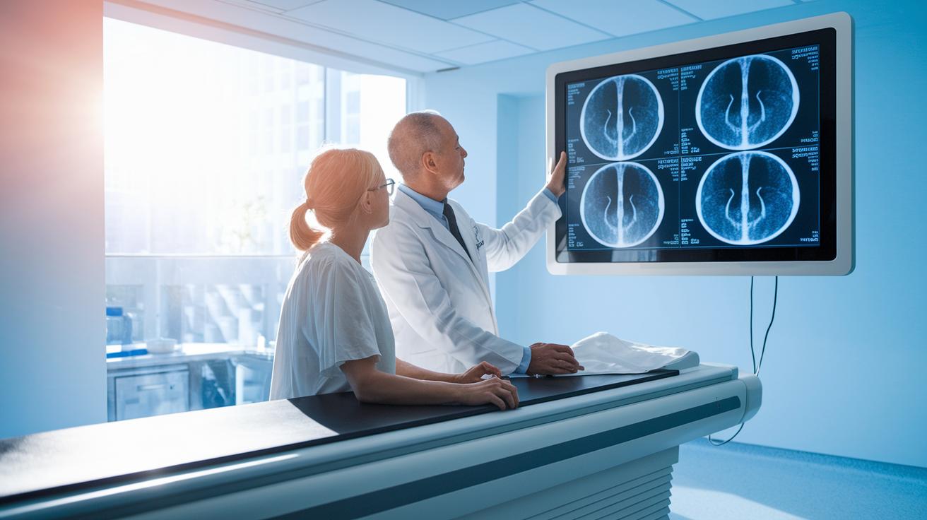
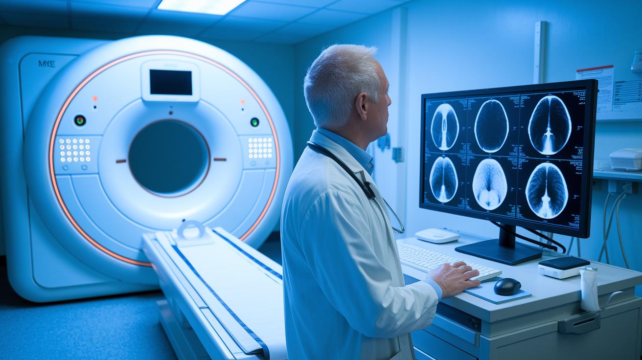
Clinical Impact of Medical Imaging Advances in Diagnosis and Care
Modern imaging tools are now a big part of everyday doctor visits. They give quick, clear pictures of what’s happening inside your body. It’s like watching a live movie of your insides, making it easier for doctors to spot problems and plan care fast.
Neurodiagnostic Applications
Doctors now use advanced MRI and PET scans to look at the brain. These scans help spot early signs of strokes and brain diseases by showing tiny differences in brain tissue. It’s like pausing a movie at a critical scene to get a closer look. This way, doctors can catch problems early and start treatment sooner.
Cardiac Imaging Breakthroughs
New methods like dynamic MRI and low-dose CT angiography let doctors see a beating heart in near real-time. These techniques capture the heart’s movements and reveal issues like blockages or irregular heartbeats. Imagine watching a highlight reel of your heart in action, this clear view helps guide quick treatment decisions.
Oncology Imaging Progress
In the fight against cancer, PET-CT scans combined with special contrast agents help find tumors at the very start. This technology mixes images of cell energy use with detailed body pictures to pinpoint small tumors. Early detection like this means treatment can begin right away, making a big difference for patients.
Interventional diagnostics and image-guided surgery add even more power to these advances. During surgery, real-time imaging helps doctors see the borders of a tumor, so they can remove cancer cells more precisely. Plus, virtual reality training has boosted surgical skills tremendously between 2016 and 2021 (impact of medical breakthroughs on healthcare quality – https://buzzyandclever.com?p=1803). All of this shows how modern imaging continues to improve patient care every day.
Emerging Research Trends and Future Directions in Medical Imaging Advances

Researchers are trying out fresh ideas that could change how we see and use medical imaging. They’re exploring methods that reveal not just the shape of organs but also how they work. For example, new functional imaging shows the inner workings of our organs, while high-field MRI gives us detailed maps of the brain. And there are exciting techniques that capture live movement, like the beating of your heart or the motion of your joints.
Doctors are also benefiting from computer simulations that let them practice surgeries on a virtual copy of a patient’s body. This means surgeons can plan and prepare for tricky procedures using a detailed model. In addition, scientists are testing improved contrast agents (special substances that make images clearer) so that images come out with greater precision.
- Functional MRI improvements: Researchers are creating MRI methods that show both the structure and function of our organs. This makes it easier to spot problems early, especially in the brain.
- Real-time dynamic imaging: New tools are capturing live movements, like your heart beating or joints moving, so doctors can see exactly how your body works during activity.
- Simulation-based surgical planning: With advanced computer models that create detailed virtual replicas of a patient’s body, surgeons can practice and plan for complex surgeries.
- Quantitative biomarker imaging: Scientists are refining ways to measure biological markers (signals that tell us about tissue health) so doctors get clear, numerical data to help guide treatment.
These emerging ideas could change how we find and treat illnesses by 2030. As they develop, they promise faster, clearer images and even personalized insights that may lead to treatments tailored just for you.
Safety Standards and Regulatory Framework for Medical Imaging Advances
Medical imaging is moving forward with clear standards that help hospitals and clinics keep images safe and easy to share. One key player in this effort is the Vendor Neutral Archive (VNA). The VNA makes it possible to store and retrieve pictures from many different systems by using open standards. This means modern CT scans now use less radiation and skip some invasive steps that could cause blood clots.
Radiology safety today focuses on a few simple ideas that keep things running smoothly:
| Practice | Why It Matters |
|---|---|
| Dose Tracking | Constantly monitoring radiation to keep patient exposure low. |
| Equipment Calibration | Regular checks ensure machines work accurately and reliably. |
| Workflow Quality Assurance | Standard methods keep imaging secure and efficient. |
Looking ahead, new imaging tools and smarter software will add some challenges. Regulators are likely to update rules with ideas like open source imaging tools to handle these changes. This fresh approach could bring more flexibility and help safety rules keep up with technology while making sure patients always get the best care.
Final Words
In the action, we saw modern medical imaging advances shaping diagnostics and patient care. We explored how upgrades in imaging systems, AI assistance, and improved hardware and software boost clarity and speed in scans.
These breakthroughs set the stage for safer, faster, and more precise practices. Moving forward, medical imaging advances hold the promise of transforming diagnoses and making healthcare better for everyone.
FAQ
What does medical imaging in biomedical engineering involve?
The role of medical imaging in biomedical engineering involves using tools like X-rays, MRI, and ultrasound to study body structures and support clinical research.
What is NIH imaging and why is it important?
NIH imaging refers to research funded by the National Institutes of Health that improves imaging methods for better diagnostic accuracy and enhanced patient care.
How do advances in medical research impact imaging techniques?
Advances in medical research boost imaging techniques by supporting quicker image sharing and more precise diagnostics, leading to improved clinical outcomes.
What are some findings in the latest medical research regarding imaging?
The latest research findings in imaging include breakthroughs in digital mammography, minimally invasive CT protocols, and AI-driven image analysis, which all speed up diagnosis and treatment.
What is the role of the National Institute of Biomedical Imaging and Bioengineering?
The National Institute of Biomedical Imaging and Bioengineering funds work that pushes imaging technology forward, merging clinical needs with innovative engineering solutions.
How do NIH-funded breakthroughs affect imaging technology?
NIH-funded breakthroughs drive the development of advanced digital radiography and imaging protocols, contributing to faster, more accurate diagnostic processes.
What technology is used in radiology for imaging advancements?
Technology used in radiology includes digital radiography, CT, MRI, ultrasound, and AI algorithms, all of which work together to improve diagnosis speed and accuracy.
