Have you ever wondered what secrets your brain can reveal without any cuts? Modern brain scans let us see live neural activity, almost like watching a movie instead of looking at a picture. Now, doctors can pick up on tiny signals that suggest mental health issues even before any clear symptoms appear. This fresh approach gives healthcare a new tool to tailor treatments and react quickly to early signs. Stick with us as we dive into how this clever technology is changing the way we look at mental health.
Breakthrough Non-Invasive Brain Imaging Techniques Transforming Mental Health Diagnostics
Lately, brain imaging technology has made some amazing strides. New methods let us explore the brain’s structure and its activity without any invasive procedures. This means doctors can now see subtle changes in real time and get a clear idea of what’s going on, whether it’s depression, trauma, or early signs of cognitive decline.
Instead of sticking with old scans that capture just a still image, modern tools capture the brain in action. Imagine watching a movie rather than looking at a photo. This dynamic view allows for more precise evaluations and helps set the stage for early intervention and personalized treatment plans.
Here are some of the standout techniques making a big difference today:
- fMRI: This method offers a live map of brain activity along with detailed images of its structure. It helps pinpoint exactly where something might be off.
- EEG: By recording fast electrical signals from the brain, EEG lets doctors follow neural events as they happen.
- PET: This technique focuses on how the brain uses energy by employing tiny, safe markers (radiotracers) to show active biochemical changes.
- SPECT: By checking blood flow in different regions of the brain, SPECT picks up on functional patterns that static images might miss.
By combining these advanced methods, health professionals can detect issues early and create treatment plans that truly suit each individual. It’s a major step toward smarter, more personalized mental health care, and it’s pretty exciting to see science work in such a tangible way.
fMRI and EEG Innovations Driving Advanced Neural Mapping in Mental Health Diagnostics
fMRI and EEG are changing how we look at mental health. They let us see what the brain is doing right away, so doctors can find hidden signals early and offer care that fits each person.
Functional MRI Innovations
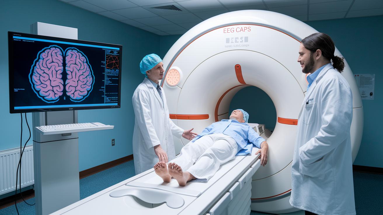
New fMRI machines work fast and use smart computer programs to create live maps of the brain. This means doctors can watch which parts of the brain light up as things happen. The computer tools help spot tiny changes that might hint at conditions like epilepsy or Alzheimer’s. By mixing detailed pictures with live data, we get a clearer view of our brain in everyday life, all without any invasive procedures.
EEG Advancements
Recent advances in EEG include sensor caps you can wear that pick up electrical signals from the brain with great clarity. They reduce background noise and use smart event detection to quickly flag anything unusual. Because these devices are portable and reliable, they let us monitor brain activity in different settings, making them a practical tool for tracking mental health.
By combining data from both fMRI and EEG, doctors can fine-tune diagnoses and keep a close eye on treatment progress. This blend of technologies helps bring personalized care and early intervention to mental health diagnostics.
Precision Diagnostic Scanning: PET vs SPECT in Noninvasive Brain Assessment for Psychiatry
PET and SPECT give us different ways to see how the brain works. PET uses radiotracers (special chemicals that light up metabolism) so doctors can spot areas that use energy in an unusual way. This might point to conditions like Alzheimer’s or brain tumors. SPECT, meanwhile, looks at blood flow in the brain and can help reveal changes linked to depression, PTSD, or injuries from accidents.
PET scans give very clear, detailed images, but they usually take longer. SPECT is faster and has stood the test of time with over a decade of research backing it up. In many cases, doctors blend SPECT with MRI and CT scans to get a full picture of both the brain’s structure and its function. This approach balances the deep insights into cellular activity with quick checks on blood circulation, refining how we diagnose psychiatric conditions.
| Modality | Diagnostic Focus | Resolution & Speed | Use Cases |
|---|---|---|---|
| PET | Metabolic activity | High resolution, longer scan time | Alzheimer’s, tumors |
| SPECT | Blood flow patterns | Moderate resolution, rapid acquisition | Depression, TBI |
Using these scanning techniques helps doctors design treatment plans that match each patient’s unique brain profile, making care more personal and effective.
Comparative Advantages of Breakthrough Brain Imaging Over Traditional MRI and CT

Advanced imaging methods like fMRI and EEG give us a richer view of the brain. They show not only the structure but also how the brain works in real time. Traditional MRI gives clear images of brain anatomy, but it can’t capture the brain's activity at any given moment. CT scans are fast but only provide still photos, and they expose patients to radiation. It’s like having a detailed map versus watching live traffic, one shows where everything is, and the other shows how things are moving.
Breakthrough imaging techniques also make the experience safer and more comfortable for patients. These methods do not use radiation, so they lower the risk of side effects. Plus, they remove the need for contrast agents that some patients might not like. With fewer risks, patients feel more at ease during the scan, and the process is easier for everyone.
New speed improvements and smart, AI-driven tools are changing how quickly and accurately doctors can spot brain issues. Automated systems review imaging in a flash, helping clinicians notice tiny changes early. This mix of fast scans and clever algorithms speeds up diagnosis and offers better hints about what might come next, ensuring that care is prompt and tailored to each patient's needs.
Clinical Relevance and Case Studies Showcasing Non-Invasive Neurodiagnostic Innovation
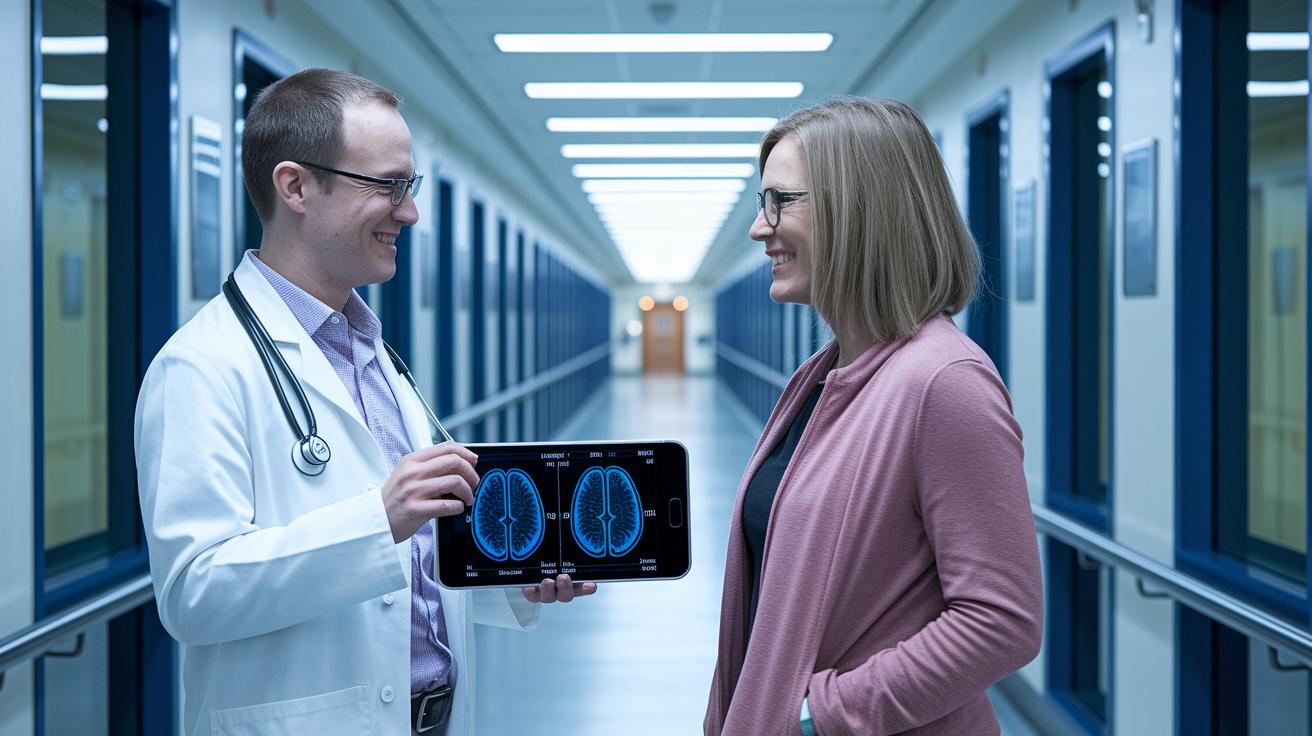
Many mental health clinics are now using gentle imaging methods to check how the brain is working. These clinics even offer same-day scans, so it all fits easily into everyday routines. Research shows that fMRI (a scan that pictures brain activity) matches up well with tools that measure depression levels. At the same time, SPECT scans (which track blood flow in the brain) reveal changes linked to issues like OCD and anxiety. These findings help doctors tailor treatments and keep a close eye on progress, showing just how promising advanced brain tests can be in mental health care.
fMRI in Depression Diagnosis
Recent studies have used fMRI to map out brain activity in people with depression. The results are clear: the scans pick up small changes in the brain areas that manage mood. Patients with distinct brain patterns often see real improvements after getting treatments customized just for them. This solid information helps clinicians fine-tune their methods and makes the connection between brain scans and everyday results even stronger.
SPECT in Anxiety Disorders
SPECT imaging has given us a straightforward look at how blood moves in the brains of people dealing with anxiety. Doctors have spotted odd blood flow patterns that often lead them to adjust treatments immediately. Later scans usually confirm that these tweaks are working by showing better blood flow in the targeted brain areas. Efforts continue to make these scanning methods more uniform and available in more clinics, so diagnosis and treatment can get even better over time.
Future Directions in Innovative Non-Invasive Brain Imaging for Mental Health Diagnostics
AI and machine learning are changing how we look at images. These clever programs catch tiny changes in brain activity that we might miss otherwise. They help doctors spot small differences early on so they can plan personalized care sooner. For example, these systems now pick up little shifts that might be early signs of brain conditions.
Portable imaging devices are making it easier to get brain scans wherever needed. Imagine a gadget that mixes the best parts of fMRI (a scan that shows brain structure) and EEG (a test that checks brain waves) into one. Soon, patients might get advanced brain scans outside traditional hospitals, making regular check-ups a simple part of daily life.
Researchers are also working on combining different types of scans into one machine that can capture the brain's structure, energy use, and electrical signals all at once. With smart models being developed to predict how conditions might progress, these new systems could help tailor treatments even better. All these breakthroughs hint at an exciting change in how we diagnose mental health issues.
Final Words
In the action, we explored advanced imaging techniques that bring fresh clarity to brain health. From the real-time maps of fMRI to the electrical monitoring of EEG, and the metabolic insights from PET and SPECT, the blog highlights how these techniques are making mental wellness evaluations more accessible and precise.
Modern methods now outshine traditional imaging by making assessments faster and safer. Breakthrough non-invasive brain imaging for mental health diagnostics is paving the way for early detection and custom care. Embrace the positive changes now!
FAQ
What does electrical stimulation of the brain tell us about psychology?
The electrical stimulation of the brain informs psychology by showing how neural circuits react to targeted inputs. It offers insights into mood regulation and cognitive processes.
What is the Alzheimer’s breakthrough predicted for 2025?
The Alzheimer’s breakthrough in 2025 signifies emerging methods for earlier detection and improved treatment. It aims to shift how we approach patient care and management of Alzheimer’s symptoms.
What is a brain stimulation test?
The brain stimulation test evaluates how specific brain regions respond to controlled electrical inputs. It helps clinicians understand neural activity patterns for more precise diagnosis.
What are common brain stimulation techniques?
The common brain stimulation techniques involve non-invasive methods, such as magnetic and electrical stimulation, which adjust neural activity to address condition-specific symptoms.
What does ultrasound for Alzheimer’s cover on 60 Minutes?
Ultrasound for Alzheimer’s, featured on 60 Minutes, demonstrates how sound waves can be used non-invasively to detect brain changes, potentially leading to earlier intervention strategies.
How does brain stimulation therapy help with depression?
Brain stimulation therapy helps with depression by using controlled impulses to adjust brain activity in targeted regions. This technique can pave the way for symptom relief where other treatments may fall short.
What is a brain stimulation device and its role?
A brain stimulation device delivers precise magnetic or electrical pulses to specific brain areas. It plays a key role in both research and treatment by modifying neural functions.
How can brain stimulation therapy be administered at home?
Brain stimulation therapy at home involves portable systems designed for safe, controlled use outside clinical settings. These emerging devices extend treatment accessibility while maintaining treatment efficacy.
