Have you ever wondered about the secret details hidden inside your body that only special imaging tools can reveal? Today, brilliant new technology lets doctors see what's going on inside you with a speed and clarity that feels almost magical. Smart computer programs (algorithms that help make decisions) and lifelike 3D images work together like a well-timed duo, catching issues earlier and speeding up care. It’s like switching on a bright light in a dark room, suddenly, everything becomes clear. Let’s explore these cool developments in medical imaging and see how they help doctors give faster and more precise care.
Key Breakthrough Technologies in Medical Imaging Diagnostics
Medical imaging diagnostics is a safe and smart way to make pictures of the inside of the body without any cutting. Doctors use these images to quickly figure out what might be wrong and decide on the best treatment. It’s a big help in both everyday check-ups and sudden emergencies.
New imaging tools are changing the game for doctors as they watch and treat health concerns. Five key ideas are driving this change. These ideas mix smart computer programs with fresh ways to see images, making it easier for doctors to get clear and fast results. For more details, check out Medical technology breakthroughs.
- Artificial Intelligence and Machine Learning in radiology – smart computer programs that learn and help spot issues on images.
- Advanced visualization with 3D rendering, VR/AR – cool tools that let you see the body in 3D or even use virtual reality to explore complex parts.
- Web-based enterprise imaging systems (Vendor Neutral Archives) – systems that let doctors pull up images on any device to work together easily.
- Off-site cloud storage for image management – secure online storage that keeps your images safe and easy to access.
- AI-driven medical image data management with universal ontologies – clever systems that organize data from different sources so everything works together smoothly.
These new technologies speed up how quickly and accurately doctors can diagnose problems. By automating routine tasks and showing clear, high-quality images, they cut down the time between scanning and treatment. This progress helps ensure that patients get the expert care they need, right when they need it.
AI-Powered Radiology Tools Transforming Diagnostic Imaging
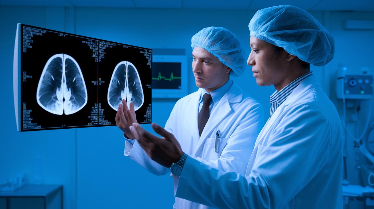
Artificial intelligence and machine learning are changing how radiologists work. These smart tools scan heaps of images in real time to spot important problems quickly. They use automated programs (these are computer systems that work on their own) to notice details that might be missed during routine checks. This means radiologists can get help from technology that finds hidden patterns and speeds up the review process.
Take Aidoc, for example. It can detect a brain bleed on a CT scan almost as soon as it happens, so urgent cases are noticed right away. Then there's Arterys, which uses cloud-based AI to review heart and lung scans, cutting down the time between taking a picture and making a diagnosis. Viz.ai works with brain scans to flag areas that might indicate a stroke, and Rayscape looks at chest images to catch early signs of issues. Each of these tools gives radiologists a boost, helping them work both faster and more accurately.
Siemens, on the other hand, has an AI tool for chest CT scans that measures bones and marks suspicious spots, making it easier to trust the results. Philips has improved its MRI process so a lumbar scan now takes under one minute instead of over three. By speeding things up and automating routine tasks, these technologies keep images clear and reduce the need for repeated scans. All of this helps doctors diagnose patients faster and with more confidence.
These AI tools act like a steady, watchful assistant, taking care of everyday jobs so radiologists can focus on tricky cases. That means patients get prompt, precise diagnoses and treatments that fit their needs. Isn't it amazing how technology can help make healthcare smoother and quicker?
Next-Gen MRI and CT Innovations in Medical Diagnostics
Have you ever wondered how far medical imaging has come? In 1895, simple X-rays first let doctors see inside the body. By the 1970s, CT scans built on those early ideas, offering clearer, more detailed pictures. Then came MRI, which uses strong magnets and radio waves (invisible signals that help create pictures) to capture amazing details. These innovations paved the way for even smarter imaging techniques today.
In the 2000s, exciting methods like fMRI and diffusion tensor imaging started gaining attention. fMRI shows brain activity by tracking changes in blood flow, while diffusion imaging follows water movement through tissues, revealing nerve pathways. CT scans also got a big boost from new methods like iterative reconstruction and dual-energy imaging, making scans super fast and detailed. In some cases, advanced CT scanners complete scans in less than 500 milliseconds, especially important when every tick counts during a heart exam.
These advancements mean patients enjoy quicker scan times and clearer images. Better images help doctors cut down on repeat scans and spot problems with ease. With less blurring and distortion, radiologists can make confident, fast diagnoses, ultimately leading to better patient care.
Molecular and Functional Imaging Innovations for Precise Diagnostics
Molecular imaging uses special contrast agents that attach to cells and let us see tiny processes happening inside the body. New types of these agents lock onto specific markers, helping us map diseases accurately. This approach not only shows body structures but also reveals the chemical activities going on in tissues, giving doctors clues for fast and precise diagnoses.
One great example is FDG-PET. This technique tracks how the body uses sugar, lighting up areas with high activity, often where cancer cells are found. It works by using a small amount of radioactive material that acts like a spotlight on unusual tissues. New metabolic tracers now give even more detail about tissue function. This means doctors can spot problems earlier, even before symptoms show up, which speeds up decisions and refines treatment plans.
Functional imaging methods like task-based fMRI record real-time brain activity by tracking changes in blood flow. When we mix data from both anatomical and metabolic scans, such as with hybrid PET/CT or PET/MRI systems, we get a full picture of a patient’s condition. This combined view helps in evaluating complex conditions from many angles while streamlining the process by replacing multiple tests with one clear scan, ultimately leading to more personalized and timely care.
Cloud-Based and Mobile Platforms Enhancing Medical Imaging Diagnostics
Cloud PACS does so much more than just store images digitally. It gives you flexible storage, strong security measures, and a pay-as-you-go model that works well for handling huge image collections. This means doctors can see images from anywhere, letting them make quick decisions even when they're not in the hospital. Plus, it grows with your needs and helps cut down on expensive onsite equipment.
Imaging APIs are now making it easier for different systems to "talk" to each other by standardizing the way DICOM (a common format for medical images) information is shared. They help automate everyday tasks, which cuts down on manual work and keeps processes smooth. By linking things like electronic health records and radiology systems, these APIs make sure patient data is current and easy to get to. This clear, automatic transfer of data means fewer holdups and better integration of diagnostic tools.
Mobile imaging apps are turning point-of-care scanning on its head by bringing diagnostic tools right to the patient’s bedside. They let clinicians capture and check ultrasound images and other scans in real time, cutting down the wait for a specialist’s opinion. This quick access is super important in emergencies or in far-off areas where every minute counts. With these mobile solutions, doctors have a handy, on-the-go way to keep patient care moving fast.
Vendor Neutral Archives make it simple for clinicians to get images and reports from different systems and locations. This easy access boosts collaboration among specialists and supports things like tele-radiology, where experts review images from a distance. By helping everyone work together better, VNAs play a key role in speeding up patient diagnoses and care.
Future Directions in Medical Imaging Diagnostics: 3D, Telemedicine, and Beyond

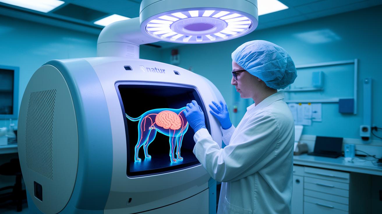
We're stepping into a whole new era for looking inside the human body. Imagine using 3D rendering (making images that pop out in three dimensions), virtual reality (an immersive digital world), and augmented reality (overlaying computer images onto what you see in real life) to get a near-touchable view of our inner workings. These advances give doctors a lifelike look at complicated body systems and help them plan surgeries more confidently. And with techniques like virtual biopsy, where images stand in for traditional, more invasive tests, the diagnostic process becomes gentler for patients.
Here are some exciting ways this technology is transforming care:
- 3D reconstructed models that guide surgeons during operations
- Image-based virtual biopsies that reduce the need for invasive sampling
- AI-driven predictive tools (smart computer programs that learn from data) for spotting issues early
- Wearable scanning devices for ongoing health checks
- Integrated telemedicine dashboards that let doctors review images and results from afar
By blending these cutting-edge tools, healthcare teams can shape diagnostic paths that truly cater to each patient. With patient portals and telemedicine (remote consultations via the internet), doctors and patients can explore scans and discuss findings without being in the same room. Meanwhile, AI systems sift through vast amounts of data to alert us to potential health concerns before they become serious. This mix of immersive visuals, constant remote access, and clever data analysis is laying the groundwork for care that’s not only faster and more accurate but also more personal. As these trends continue to evolve, diagnostic processes will get even more tailored, streamlined, and interconnected, ushering in a future where every patient gets a care experience made just for them.
Final Words
In the action-packed article, we reviewed how modern imaging tools are reshaping patient care, from AI-powered radiology and next-gen MRI/CT to molecular imaging and innovative cloud-based platforms. Each breakthrough lights the way for faster, clearer diagnoses and improved treatment plans.
This exploration reminds us that medical imaging innovations: breakthrough technologies in diagnostics are enhancing daily healthcare. Keep an eye on these evolving trends and cherish the inspiring progress that makes science more accessible and impactful for us all.
FAQ
What is medical imaging and diagnosis?
The term “medical imaging and diagnosis” means using non-invasive techniques—such as MRI, CT, ultrasound, and X-rays—to create body images that help doctors detect conditions and plan treatments quickly.
What are the main types of diagnostic imaging techniques?
The main diagnostic imaging types include X-rays, computed tomography (CT), magnetic resonance imaging (MRI), and ultrasound, each providing different views of internal structures for accurate assessments.
What are common imaging techniques and examples of imaging technology?
Common imaging techniques cover X-rays, CT scans, MRIs, ultrasounds, and nuclear imaging. These methods offer detailed insights into body parts, supporting doctors in making effective diagnoses.
What are the benefits of medical imaging?
The benefits of medical imaging include faster detection of issues, more accurate treatment planning, a non-invasive approach, and overall improved outcomes in patient care and recovery.
What information is typically found in a medical imaging modalities PDF?
A medical imaging modalities PDF typically explains different imaging methods, describes how each works, outlines their clinical uses, and provides technical details, serving as a handy guide for medical professionals.
What are the latest advanced imaging technologies and new radiology tools?
The latest imaging technologies use artificial intelligence, enhanced 3D visualization, and cloud-based platforms. These tools make scans faster and more accurate, helping radiologists deliver better, patient-focused care.
