
Have you ever thought that a little gadget could change the way we handle emergencies? Today’s point-of-care ultrasound (PoCUS, which is a small machine that shows live images) lets doctors see key details right at a patient’s side. Instead of using big, clunky machines, doctors now have a handy, portable tool that helps them make quick decisions when every second counts.
This clever technology not only speeds up how fast a diagnosis can be made but also helps doctors spot hidden injuries more accurately. In really critical moments, having such a tool can make all the difference in saving a life.
How breakthrough point-of-care ultrasound reshapes emergency diagnostics
Point-of-care ultrasound (PoCUS) gives doctors the key information they need in a flash, especially when every second counts. Since the 1990s, these real-time imaging tools (they show live visuals) have grown into essential devices that help make fast decisions during emergencies. Using PoCUS right at the patient’s bedside has changed how we handle trauma and other urgent cases.
Today’s PoCUS machines are built to be affordable, compact, and easy to use in bustling emergency rooms. Early portable units made it possible to perform FAST exams (quick checks for internal injuries) which cut down the wait time for diagnoses and let doctors get critical details in moments.
- Rapid diagnosis leading to life-saving actions
- Sharper accuracy when spotting internal injuries
- Portability that makes bedside evaluations a breeze
- Live imaging that helps make speedy care choices
- Smoother routines that boost overall patient care
Unlike older imaging machines that were big and fixed in radiology rooms, these breakthrough PoCUS systems provide instant insights right where patients are. In the past, moving patients to scan machines could delay treatment. Now, modern PoCUS lets medical teams do accurate checks on the spot, improving outcomes and helping everyone get the care they need faster.
Historical evolution of point-of-care ultrasound breakthroughs in emergency care

Ultrasound has come a long way. What started as a clunky tool kept in a very specific room has transformed into a handy device that almost fits in your coat pocket, delivering live images right at the bedside.
1960s–1980s: Emergence of emergency imaging
Back in the day, in the 1960s through the 1980s, ultrasound machines were enormous and stuck in radiology departments. They were so bulky that taking a patient to get scanned was completely normal. Can you imagine that? Moving a patient for a scan was just how things were done!
1990s: FAST exams go bedside
Then came a major change in the 1990s. Portable ultrasound devices made it possible to run FAST exams (quick trauma scans) where patients actually needed them, right by the bedside. This was revolutionary because it meant doctors could spot serious internal injuries right away, without the hassle of dragging a patient across the hospital.
2000s: Formalized training and guidelines
By the 2000s, teaching hospitals and major emergency medicine groups started to include ultrasound training as part of residency programs. This ensured that more doctors could skillfully operate these machines. Picture a resident learning how to use a live scanner during training, it really set the stage for faster, more accurate diagnoses.
2010s–present: Miniaturization and tele-ultrasound
Today, things have gotten even cooler. Ultrasound devices have shrunk to tiny sizes and now come with tele-ultrasound features. This lets remote experts help doctors interpret images in real time, even in areas that once had no access to such advanced technology. It’s amazing to think how far we’ve come!
Each of these decades built on previous breakthroughs, leading us to today’s powerful and accessible tools that help emergency care teams make quick, informed decisions.
Breakthrough features of modern portable imaging devices for emergency care
Modern portable imaging devices are changing how emergency care works. These handy tools pack advanced ultrasound (a way to see inside the body using sound waves) into a tiny unit that can easily fit into a coat pocket. They mix small, efficient hardware with smart software that helps doctors quickly mark up images and save them in DICOM format (a common system for storing medical images). Plus, wireless probe connections keep things clean and fast, and tele-ultrasound features let specialists offer advice from afar. Even better, because they're made cheaply, these devices can be used in places with fewer resources, speeding up emergency care a lot.
| Feature | Description | Emergency Benefit |
|---|---|---|
| Miniaturized Design | Compact hardware that fits in a coat pocket | Makes it super easy to move and use quickly |
| Integrated Software | Makes image marking and saving in DICOM simple | Helps doctors get the right information fast |
| Wireless Probe Connectivity | No messy wires, keeping everything neat and safe | Helps speed up emergency procedures |
| Tele-Ultrasound | Lets doctors talk to specialists remotely | Provides expert help when it’s needed most |
| Affordable Manufacturing | Made using low-cost methods | Allows use in low-resource areas |
These amazing tech improvements mean that doctors can check patients quickly right where they are. Every feature is built to bring key information right to the hands of emergency teams, helping them give better care and make life-saving choices on the spot.
Clinical performance of novel point-of-care ultrasound in acute trauma diagnostics
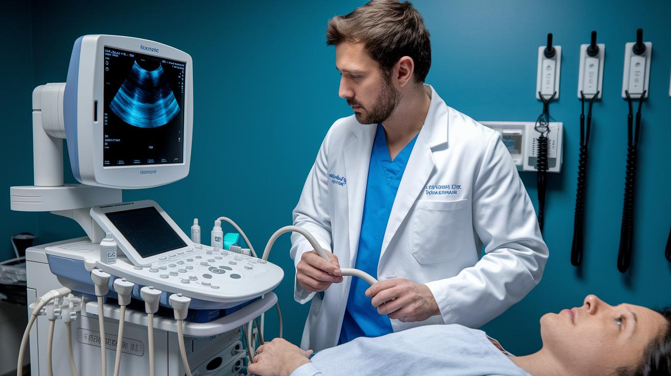
Doctors often trust FAST (a quick scan to check for internal bleeding) in emergencies. But now, they’re finding that adding an advanced lung ultrasound gives a fuller picture. In real-life cases, doctors have discovered hidden chest injuries when they used both methods together. It’s like looking at two pieces of a puzzle that, when combined, show the complete picture.
Lung ultrasound has been getting a lot of attention lately, especially thanks to what's called the B-line artifact (bright lines on the scan that show fluid in the lungs). In one study, patients with suspected lung problems had subtle signs that a FAST exam might miss, but lung ultrasound caught them. Imagine an emergency patient with vague breathing issues. The FAST exam didn’t show much, but clear B-line patterns led to quick, targeted treatment.
When you compare these two point-of-care ultrasound techniques, each has its own strengths. FAST is great for rapidly spotting internal bleeding, while lung ultrasound, with its focus on B-line markers, gives extra clues about lung problems. Recent data from clinics show that using both methods together helps emergency teams make quicker and more exact decisions when treating trauma.
Integrating cutting-edge PoCUS devices: training and implementation in emergency departments
Over the last twenty years, emergency rooms have started using PoCUS (Point-of-Care Ultrasound; a small, handy ultrasound machine used right at the bedside) as a key tool in urgent care. Based on guidelines from the 2000s by ACEP (American College of Emergency Physicians), hospitals now make sure residents learn how to use these devices. Doctors get practical training that covers everything from basic operation to safety and reading live images. This hands-on learning helps every clinician feel confident, even when time is tight.
Today, the focus has shifted to proving real skill rather than just spending time with a tool. Instead of meeting time-based requirements, medical staff must show they can effectively use PoCUS by interpreting live images and making fast, correct decisions. This modern approach not only keeps patients safe but also builds trust among team members during chaotic moments.
Hospitals are also rethinking how they set up their spaces to work smoothly with these advanced devices. They adjust room layouts and move to digital records to fit better with mobile diagnostic tools. Teams collaborate to create workspaces where PoCUS is practical and efficient. Plus, regular refresher courses keep everyone updated with the latest software improvements and new device features. All these steps ensure that even during high-pressure situations, point-of-care imaging works seamlessly, helping doctors provide quick and accurate care.
Future perspectives on next-generation breakthrough ultrasound platforms in emergency care

Tele-ultrasound (ultrasound exams done remotely using telecommunication) is changing how people get expert care. Imagine a small, rural clinic where a doctor can send live images, captured by a tiny, ultraportable scanner that fits in your pocket, straight to a specialist. This secure wireless link means doctors in areas with few resources can quickly get help from experts, which can make a huge difference in emergencies. Did you know a remote emergency room in a small town can now instantly connect with a top specialist, all thanks to tele-ultrasound technology?
Looking forward, future improvements will focus on advanced digital diagnostics and imaging boosted by AI (artificial intelligence, which helps computers think a bit like humans). Researchers are creating new tools that offer more accurate and real-time decision support. As these devices get even smaller and their batteries last longer, emergency teams can have reliable, innovative tools wherever they are needed. This progress in point-of-service imaging promises faster and sharper assessments, which is vital when every second counts in emergency care.
Final Words
In the action, our discussion showcased the evolution of emergency diagnostics, from early bulky devices to today’s cutting-edge portable units. We explored how rapid diagnostic scanning and real-time imaging improved patient care and stressed the benefits of breakthrough point-of-care ultrasound technology in emergency care.
By highlighting clinical impacts and training advancements, this piece paints a clear picture of how modern ultrasound reshapes emergency evaluations. The future looks bright as these innovations continue to refine and speed up life-saving decisions.
FAQ
Q: What is the cost of a point-of-care ultrasound machine?
A: The cost of a point-of-care ultrasound machine varies based on features, brand, and clinical setting. It offers affordability combined with rapid diagnostic scanning for emergency care.
Q: What do Philips Flash Ultrasound systems offer?
A: The Philips Flash Ultrasound systems offer quick imaging solutions with real-time diagnostic capabilities, benefiting clinical workflows in fast-paced emergency environments.
Q: What is the history of point-of-care ultrasound?
A: The history of point-of-care ultrasound starts in the 1990s when portable machines enabled bedside assessments like FAST exams, which reduced delays and improved emergency diagnostics.
Q: How are current and evolving clinical concepts applied in emergency medicine?
A: Current and evolving clinical concepts in emergency medicine use advanced ultrasound techniques that support rapid decision-making and improve patient outcomes in urgent care settings.
Q: What opportunities are available in Philips Ultrasound careers?
A: Philips Ultrasound careers provide roles in developing state-of-the-art imaging devices, contributing to advancements in portable technology for emergency diagnostics.
Q: What are the features of Philips 5300 and Philips 5000 Ultrasound systems?
A: The Philips 5300 and Philips 5000 Ultrasound systems deliver high-quality imaging paired with user-friendly design, making them effective for quick assessments in emergency care.
Q: Which is known as the best POCUS ultrasound device?
A: The best POCUS ultrasound devices integrate portability, clear real-time imaging, and ease of clinical use, offering reliable performance in fast-paced emergency settings.